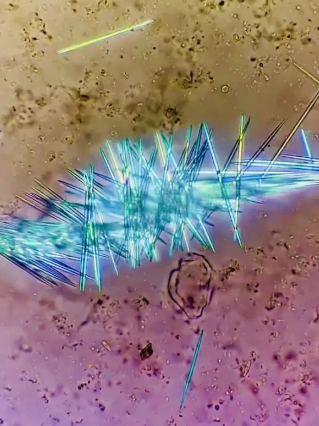
The Fascinating World Of Pineapple Anatomy: A Closer Look Under The Microscope
When you think of pineapples, the first image that comes to mind is likely its vibrant tropical exterior and juicy, sweet interior. But have you ever wondered what happens when we take a closer look at this fruit under a microscope? Pineapple under microscope reveals an entirely new dimension of this popular fruit, showcasing its intricate cellular structure and fascinating biological features. For scientists, food enthusiasts, and curious minds alike, exploring the world of pineapple anatomy provides a deeper understanding of how this fruit functions at a microscopic level. In this article, we will delve into the hidden world of pineapples and uncover the secrets hidden beneath its spiky exterior.
Understanding the structure of a pineapple is not just about satisfying curiosity; it also helps in improving agricultural practices, enhancing nutritional value, and even developing new food products. Through advanced imaging techniques and detailed analysis, researchers have uncovered the secrets of pineapple composition, giving us a clearer picture of its internal organization. This exploration opens up possibilities for innovations in agriculture, nutrition, and culinary arts. Whether you're a food scientist or simply someone who appreciates the intricacies of nature, the world of pineapple under microscope offers a captivating journey into the microcosm of this tropical delight.
As we dive deeper into this topic, we will explore the various layers of a pineapple, from its outer skin to its juicy core. By examining these layers under the microscope, we gain insight into the fruit's cellular composition, nutrient distribution, and water content. This information not only enhances our appreciation of pineapples but also contributes to advancements in food science and technology. So, let’s embark on this fascinating journey to uncover the hidden beauty of pineapples through the lens of a microscope.
Read also:Adragon De Mello Now Unveiling The Journey Of A Child Prodigy
What Can We Learn From Pineapple Under Microscope?
When scientists place a slice of pineapple under a microscope, they are greeted by a complex web of cells, fibers, and vessels that work together to sustain the fruit. The outer layer, known as the exocarp, is composed of tough, protective cells designed to shield the inner flesh from external threats. Beneath this lies the mesocarp, which contains the juicy, edible portion of the pineapple. This layer is rich in parenchyma cells, which store water, sugars, and other nutrients essential for the fruit's growth and development. Examining these structures under the microscope provides valuable insights into how pineapples thrive in their natural environment.
One of the most intriguing aspects of pineapple anatomy is its vascular system. Under the microscope, you can clearly see the xylem and phloem tissues that transport water and nutrients throughout the fruit. These vascular bundles are arranged in a specific pattern, ensuring efficient distribution of resources. By studying these structures, scientists can better understand how pineapples adapt to different growing conditions and optimize their growth processes. This knowledge can be applied to improve cultivation techniques and increase crop yields.
Why Is Studying Pineapple Under Microscope Important?
Studying pineapple under microscope is more than just a scientific curiosity; it has practical applications in agriculture and food science. For instance, understanding the cellular structure of pineapples can help farmers develop strategies to enhance fruit quality and resistance to diseases. By identifying the specific cellular components responsible for flavor, texture, and nutritional value, researchers can breed varieties that meet consumer demands. Additionally, studying the fruit's microstructure can lead to innovations in food preservation and processing, ensuring that pineapples remain fresh and nutritious for longer periods.
Another significant benefit of examining pineapple under microscope is its potential to improve sustainability in agriculture. By analyzing the fruit's cellular composition, scientists can identify ways to reduce water usage and minimize waste during cultivation. This approach not only benefits farmers but also contributes to global efforts to combat food insecurity and environmental degradation. As we continue to explore the world of pineapple anatomy, we unlock possibilities for creating a more sustainable and resilient food system.
How Does the Pineapple Under Microscope Reveal Its Nutritional Secrets?
Under the microscope, the nutritional secrets of pineapples become more apparent. The fruit's parenchyma cells are packed with vitamins, minerals, and antioxidants that contribute to its health benefits. For example, the presence of vitamin C and bromelain, a digestive enzyme, makes pineapples a popular choice for boosting immunity and aiding digestion. By studying the distribution of these nutrients within the fruit's cellular structure, scientists can better understand how pineapples provide their health benefits and how to enhance these properties through selective breeding.
Moreover, examining the pineapple under microscope allows researchers to investigate the interactions between different cellular components. For instance, how do the fruit's sugars and acids combine to create its characteristic sweet-tart flavor? Answering these questions can lead to innovations in flavor enhancement and the development of new food products. As our understanding of pineapple anatomy deepens, so too does our ability to harness its full potential for nutritional and culinary purposes.
Read also:Recent Arrests In St Lucie County A Comprehensive Overview
What Are the Key Layers of a Pineapple?
A pineapple is composed of several distinct layers, each with its own unique characteristics and functions. The outermost layer, or exocarp, serves as a protective barrier against pests and environmental stresses. Beneath this lies the mesocarp, which contains the fruit's fleshy, edible portion. This layer is rich in water, sugars, and nutrients, making it the primary source of the pineapple's flavor and aroma. Finally, the core of the pineapple consists of fibrous tissue that provides structural support and helps transport water and nutrients throughout the fruit.
By examining these layers under the microscope, scientists can gain a deeper understanding of how they contribute to the overall structure and function of the pineapple. For example, the exocarp's tough, waxy surface helps prevent water loss and protect the fruit from damage. Meanwhile, the mesocarp's dense network of parenchyma cells ensures that the fruit remains juicy and flavorful. These insights not only enhance our appreciation of pineapples but also provide valuable information for improving cultivation and processing techniques.
Can Pineapple Under Microscope Help Improve Agricultural Practices?
Absolute yes. Studying pineapple under microscope can significantly improve agricultural practices by providing insights into the fruit's growth patterns, nutrient requirements, and disease resistance. For instance, by analyzing the cellular structure of pineapples, researchers can identify the specific conditions that promote optimal growth and development. This information can be used to develop cultivation strategies that maximize yield and minimize resource usage. Furthermore, understanding the fruit's microstructure can help farmers develop strategies to combat pests and diseases, ensuring healthier and more productive crops.
In addition to enhancing cultivation practices, studying pineapple under microscope can also contribute to the development of new food products. By identifying the specific cellular components responsible for flavor, texture, and nutritional value, scientists can create innovative recipes and food items that highlight the unique qualities of pineapples. This approach not only benefits consumers but also supports the growth of the agricultural industry by creating new markets for pineapple-based products.
How Does Pineapple Under Microscope Enhance Our Understanding of Fruit Development?
Examining pineapple under microscope provides valuable insights into the processes of fruit development and maturation. By observing the changes in cellular structure and composition over time, scientists can better understand how pineapples grow and mature. For example, during the ripening process, the fruit's parenchyma cells undergo significant changes, converting stored starches into sugars and releasing aromatic compounds that enhance flavor and aroma. Studying these processes under the microscope allows researchers to identify the factors that influence fruit quality and develop strategies to optimize these characteristics.
Furthermore, examining the pineapple under microscope can reveal the mechanisms behind its unique adaptation to tropical climates. By studying the fruit's cellular structure and vascular system, scientists can identify the specific adaptations that allow pineapples to thrive in hot, humid environments. This knowledge can be applied to improve the resilience of other crops and support sustainable agricultural practices in regions with similar climates.
What Are the Practical Applications of Pineapple Microscopy?
The practical applications of pineapple microscopy extend beyond scientific research and into the realms of agriculture, nutrition, and culinary arts. For farmers, understanding the cellular structure of pineapples can lead to more efficient cultivation practices and higher yields. For nutritionists, studying the fruit's microstructure provides insights into its health benefits and how to maximize these properties. And for chefs and food scientists, examining pineapple under microscope opens up new possibilities for flavor enhancement and product development.
One of the most exciting applications of pineapple microscopy is its potential to improve food preservation techniques. By analyzing the fruit's cellular structure, researchers can identify methods to extend its shelf life and maintain its quality during storage and transportation. This approach not only benefits consumers by ensuring access to fresh, nutritious produce but also supports the global effort to reduce food waste and promote sustainability.
What Are the Challenges of Studying Pineapple Under Microscope?
While studying pineapple under microscope offers numerous benefits, it also presents several challenges. One of the primary challenges is preparing the fruit for microscopic analysis. Pineapples have a tough, fibrous exterior that can make it difficult to obtain clear, high-quality images. Additionally, the fruit's complex cellular structure requires advanced imaging techniques and specialized equipment to fully appreciate its intricacies.
Another challenge is interpreting the data obtained from microscopic analysis. The vast amount of information generated by these studies requires careful analysis and interpretation to draw meaningful conclusions. This process often involves collaboration between scientists from different disciplines, including botany, chemistry, and food science, to ensure a comprehensive understanding of pineapple anatomy and its implications for agriculture and nutrition.
What Are the Future Directions for Pineapple Microscopy Research?
As technology continues to advance, the field of pineapple microscopy research is poised to make significant strides in the coming years. Innovations in imaging techniques and data analysis methods will enable scientists to explore the fruit's microstructure in unprecedented detail, uncovering new insights into its biology and potential applications. For example, the development of non-invasive imaging techniques could allow researchers to study the fruit's internal structure without damaging it, opening up new possibilities for real-time monitoring of growth and development.
Furthermore, advancements in genetic engineering and biotechnology could lead to the creation of pineapple varieties with enhanced nutritional value, improved disease resistance, and better adaptability to changing environmental conditions. By combining these technologies with traditional microscopy techniques, scientists can create a comprehensive understanding of pineapple anatomy and its implications for agriculture, nutrition, and culinary arts.
Conclusion
In conclusion, studying pineapple under microscope offers a fascinating glimpse into the intricate world of this tropical fruit. From its protective outer layer to its juicy core, each component of the pineapple plays a vital role in its growth, development, and function. By examining these structures under the microscope, scientists can gain valuable insights into the fruit's biology and potential applications in agriculture, nutrition, and culinary arts. As we continue to explore the world of pineapple anatomy, we unlock new possibilities for improving our understanding of this beloved fruit and harnessing its full potential for the benefit of society.
Table of Contents
- What Can We Learn From Pineapple Under Microscope?
- Why Is Studying Pineapple Under Microscope Important?
- How Does the Pineapple Under Microscope Reveal Its Nutritional Secrets?
- What Are the Key Layers of a Pineapple?
- Can Pineapple Under Microscope Help Improve Agricultural Practices?
- How Does Pineapple Under Microscope Enhance Our Understanding of Fruit Development?
- What Are the Practical Applications of Pineapple Microscopy?
- What Are the Challenges of Studying Pineapple Under Microscope?
- What Are the Future Directions for Pineapple Microscopy Research?
- Conclusion


